

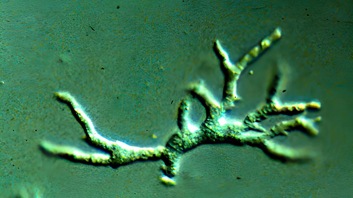
In the quiet corners of medical literature, there exist pathogens so rare that they rarely cross the threshold of a physician’s diagnostic consideration. Balamuthia mandrillaris is one such organism—a free-living, single-celled amoeba that resides in soil and water, and occasionally, with devastating consequence, in the human brain. A recent, heartbreaking case study published in BMJ Case Reports chronicles the struggle of a healthy toddler in Washington state, whose sudden decline into illness serves as a somber lesson on the dangers of diagnostic blind spots and the imperative for faster, more accurate testing in cases of neurological distress.
The Chronology of a Diagnostic Odyssey
The tragedy began with an abrupt onset of symptoms that might initially appear non-specific: vomiting, profound lethargy, and a concerning weakness on the right side of the toddler’s body. As the days progressed, the clinical picture grew more alarming. The boy developed a high fever, persistent headaches, and an increasing inability to swallow. The situation turned critical when the child experienced periods of unresponsiveness and dangerous respiratory failure, necessitating emergency hospitalization.
The Initial Misdirection
Upon admission, doctors performed a computed tomography (CT) scan, which revealed a hemorrhage in the brain, leading to the surgical placement of a shunt. Subsequent imaging of the cerebral vasculature showed narrowing and blockages—a finding that had not previously been associated with amoebic infections.
Based on this, medical teams pursued a diagnosis of childhood Takayasu arteritis, a rare, systemic inflammatory condition that affects medium and large arteries. The treatment plan involved immunosuppressive therapy. Initially, the child showed signs of improvement, and he was discharged after two weeks. However, the respite was brief. A week later, he returned to the emergency department with recurrent vomiting. Because his shunt appeared functional, he was sent home under the suspicion that his symptoms were merely side effects of his medication.
The Critical Decline
Two days later, the parents returned with their son, now suffering from fever, lethargy, and significant dysphagia (trouble swallowing). He was re-admitted, and his condition deteriorated rapidly. By day 25, he required intubation. Two days later, he became entirely unresponsive; his pupils were pinpoint, his eyes fixed upward, and he displayed extreme muscular flaccidity.
It was not until the 30th day of his medical journey—following a brain biopsy and the application of metagenomic next-generation sequencing (mNGS) on his cerebrospinal fluid—that the true culprit was identified: Balamuthia mandrillaris. By the time the pathogen was confirmed, the damage to the boy’s brain was catastrophic and irreversible. Following failed attempts at intensive antimicrobial therapy, his parents made the agonizing decision to remove his life support. An autopsy later confirmed that his brain tissue was riddled with the amoeba, accompanied by severe necrotic damage to the vasculature.
Understanding Balamuthia mandrillaris
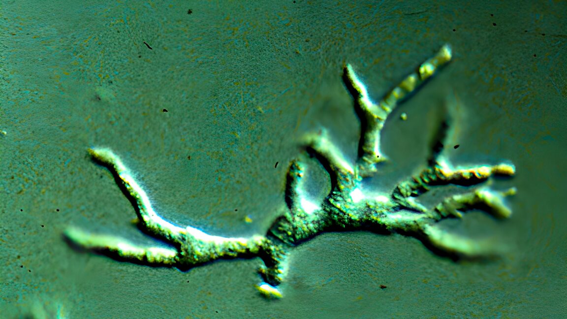
Balamuthia mandrillaris is a rare, opportunistic pathogen first identified in 1986 following the death of a mandrill at the San Diego Zoo. Since its discovery, fewer than 200 human cases have been recorded globally. Its rarity is compounded by a fatality rate that exceeds 90 percent.
Biological Characteristics and Transmission
As a free-living amoeba, B. mandrillaris thrives in soil and water. While the exact mechanism of transmission remains a subject of ongoing research, experts believe humans become infected when the amoeba enters the body through skin wounds or by inhalation of contaminated, windblown dust. Once inside, it can travel through the bloodstream to the central nervous system, where it causes amoebic granulomatous encephalitis—a severe, progressive, and often fatal infection of the brain.
Why Is It So Hard to Detect?
The challenge with Balamuthia lies in its "vague" early presentation. Symptoms such as headache, fatigue, and nausea mimic a wide array of common childhood illnesses. Furthermore, because the organism is not a common bacteria or virus, it does not show up on standard infectious disease panels. Medical teams must have a high index of suspicion to even consider testing for it, and in this specific case, the presence of vasculitis-like symptoms—which were actually manifestations of the amoeba’s damage—effectively led clinicians down the wrong path.
The Role of Immunosuppression
Perhaps the most haunting revelation in the BMJ case report is the potential role that the initial treatment plan played in the child’s decline. While immunosuppressive drugs were intended to manage the suspected Takayasu arteritis, they may have inadvertently created a more favorable environment for the amoeba to multiply.
By suppressing the body’s natural immune response, the medication may have hindered the child’s ability to fight off the invading pathogen, allowing the amoeba to ravage brain tissue more aggressively than it might have otherwise. This highlights the danger of "premature diagnostic closure"—a phenomenon where physicians settle on a diagnosis too early and ignore contradictory evidence, potentially causing more harm than good.
Lessons for Future Practice: The Path Forward
The doctors at Seattle Children’s Hospital, who authored the case report, are adamant that this tragedy should not be in vain. They have shared the details of the case with the hope of changing how similar cases are handled in the future.

Advocacy for Early Diagnostic Testing
The medical team now strongly advocates against premature diagnostic closure. They recommend that in cases of progressive vasculitis or unexplained neurological deterioration, B. mandrillaris should be included in the differential diagnosis.
Furthermore, the successful identification of the amoeba via metagenomic next-generation sequencing (mNGS) suggests a potential, less invasive way to diagnose future cases. By analyzing the genetic material within the cerebrospinal fluid, doctors can identify the "genetic signature" of the amoeba much faster than by waiting for a biopsy or relying on traditional culture methods. The team suggests that before any immunosuppressive therapy is escalated, physicians should perform comprehensive diagnostic testing for infectious microbes, including mNGS.
The Survival Blueprint
Early detection is the primary determinant of survival. The report cites a 2010 case in Kentucky, where a two-year-old boy survived the infection because it was identified within 10 days of his hospital admission. While he suffered significant neurological damage, he eventually regained the ability to hold himself upright, interact socially, and follow commands. His survival serves as a testament to the fact that while Balamuthia is formidable, it is not always a death sentence if caught in time.
Implications for Public Health and Clinical Vigilance
The story of this toddler is a stark reminder of the complexities of modern medicine, where the rarest of conditions can mimic the most common ones. It raises significant questions for clinical practice:
- Increased Vigilance: Should clinicians in warmer climates—where Balamuthia is more prevalent—be trained to consider amoebic infections for any child with unexplained, persistent neurological symptoms?
- Standardization of Testing: Should advanced genetic testing (mNGS) become a standard protocol for "diagnostic dilemmas" where traditional imaging and blood work fail to yield a clear cause?
- The Danger of Assumptions: The case serves as a warning that a "best fit" diagnosis is not the same as a confirmed one. When a patient’s condition evolves, clinicians must be prepared to pivot, reassess, and consider the "unthinkable" pathogens.
Ultimately, the parents of the young boy chose to share this story to spare other families from the same heartbreak. By illuminating the dark corners of this rare, silent invader, the medical community hopes to foster a culture of skepticism toward premature labels and an embrace of rigorous, broad-spectrum diagnostics. While Balamuthia remains an elusive and deadly threat, the path toward a future of higher survival rates begins with the recognition that even in the world of medicine, the most dangerous enemies are often the ones we fail to suspect.
